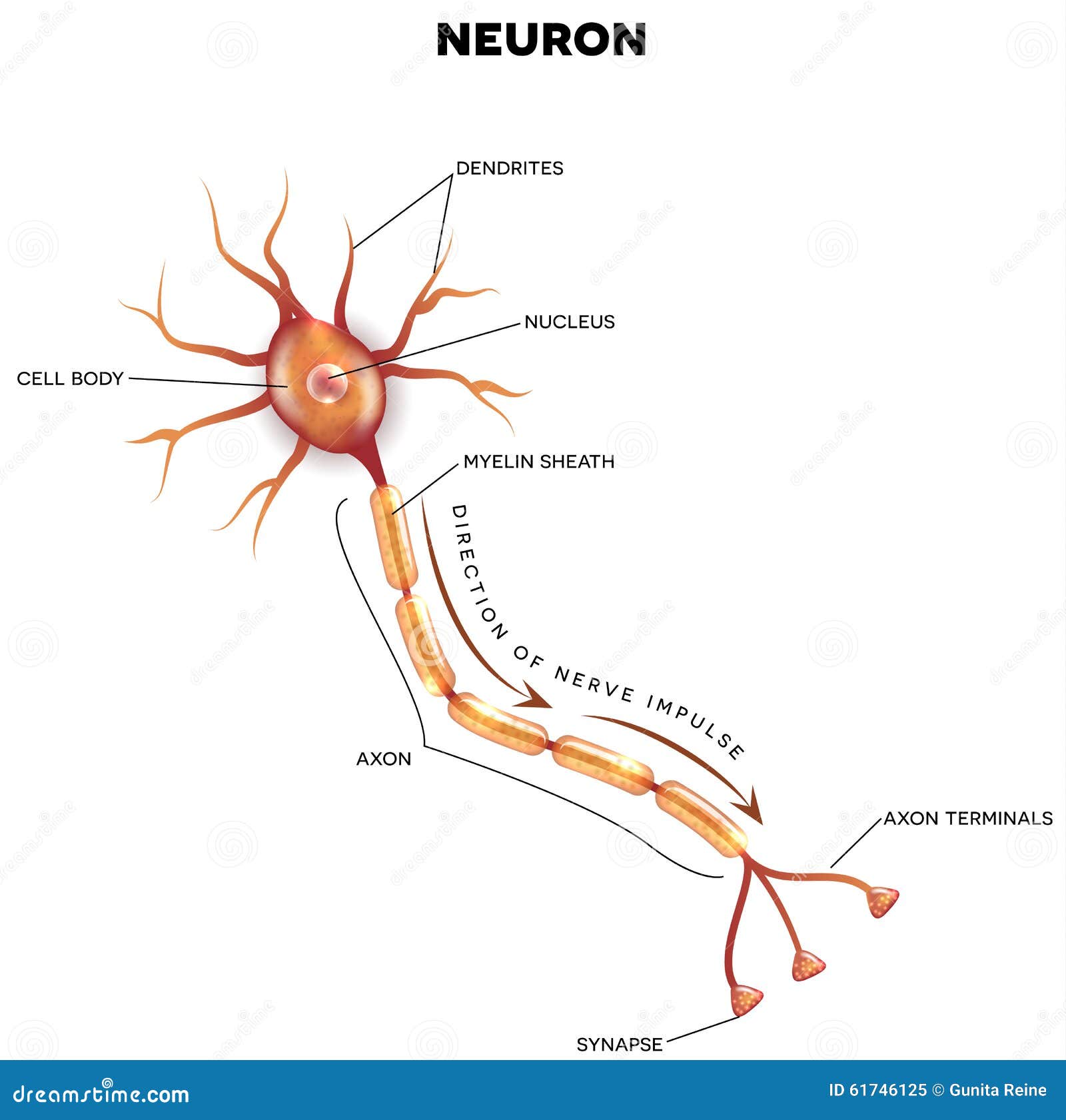
Labeled Diagram Of The Neuron Stock Vector Image 61746125
AboutTranscript. Upper motor neurons control lower motor neurons and skeletal muscle cells. Located in the cerebral cortex, these neurons follow specific pathways and their dysfunction can impact reflexes and muscle tone. Understanding their role is key to unraveling the complexities of our nervous system.

Myelinated Motor Neurons Function, Location & Types
Figure 14.28 Corticospinal Tract The major descending tract that controls skeletal muscle movements is the corticospinal tract. It is composed of two neurons, the upper motor neuron and the lower motor neuron. The upper motor neuron has its cell body in the primary motor cortex of the frontal lobe and synapses on the lower motor neuron, which is in the ventral horn of the spinal cord and.

Picture Of A Neuron Labeled Luxury Motor Neuron Detailed Accurate
Motor neurones are cells in the brain and spinal cord that allow us to move, speak, swallow and breathe by sending commands from the brain to the muscles that carry out these functions. Their nerve fibers are the longest in the body, a single axon can stretch from the base of the spinal cord all the way to the toes.
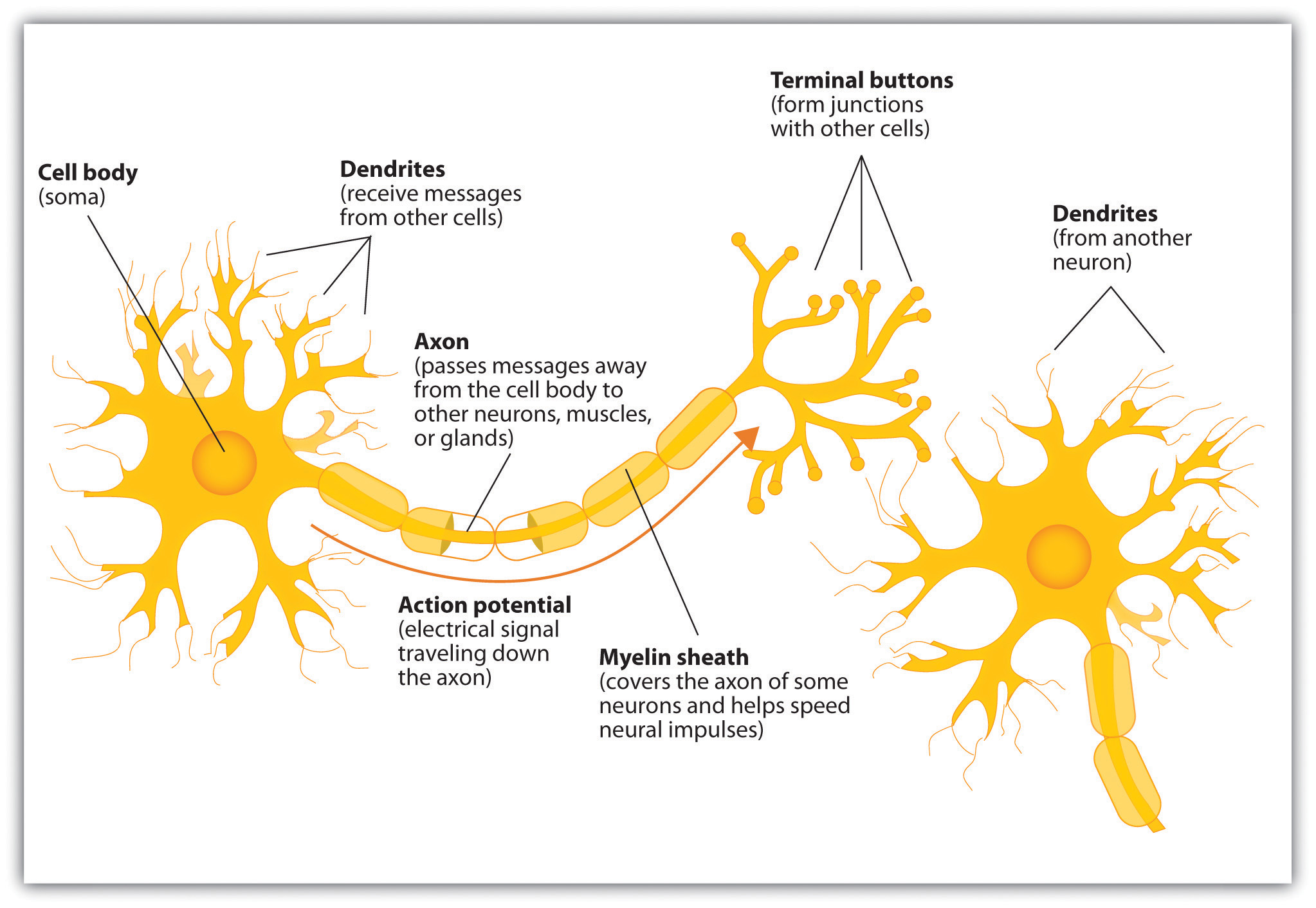
The Neuron Is the Building Block of the Nervous System USE ME
Motor neurons, also known as efferent neurons, are nerve cells responsible for carrying central nervous system signals towards muscles to cause voluntary or involuntary movement through the innervation of effector muscles and glands. Their nerve fibers are considered to be the longest in the human body .
Neurones Anjung Sains Makmal 3
Motor neurons (also referred to as efferent neurons) are the nerve cells responsible for carrying signals away from the central nervous system towards muscles to cause movement. They release neurotransmitters to trigger responses leading to muscle movement.

Neuron Diagram Straight from a Scientist
Motor neurons get information from other neurons and convey commands to your muscles, organs and glands.
:max_bytes(150000):strip_icc()/neuron-anatomy-58530ffe3df78ce2c34a7350.jpg)
Neuron Anatomy, Nerve Impulses, and Classifications
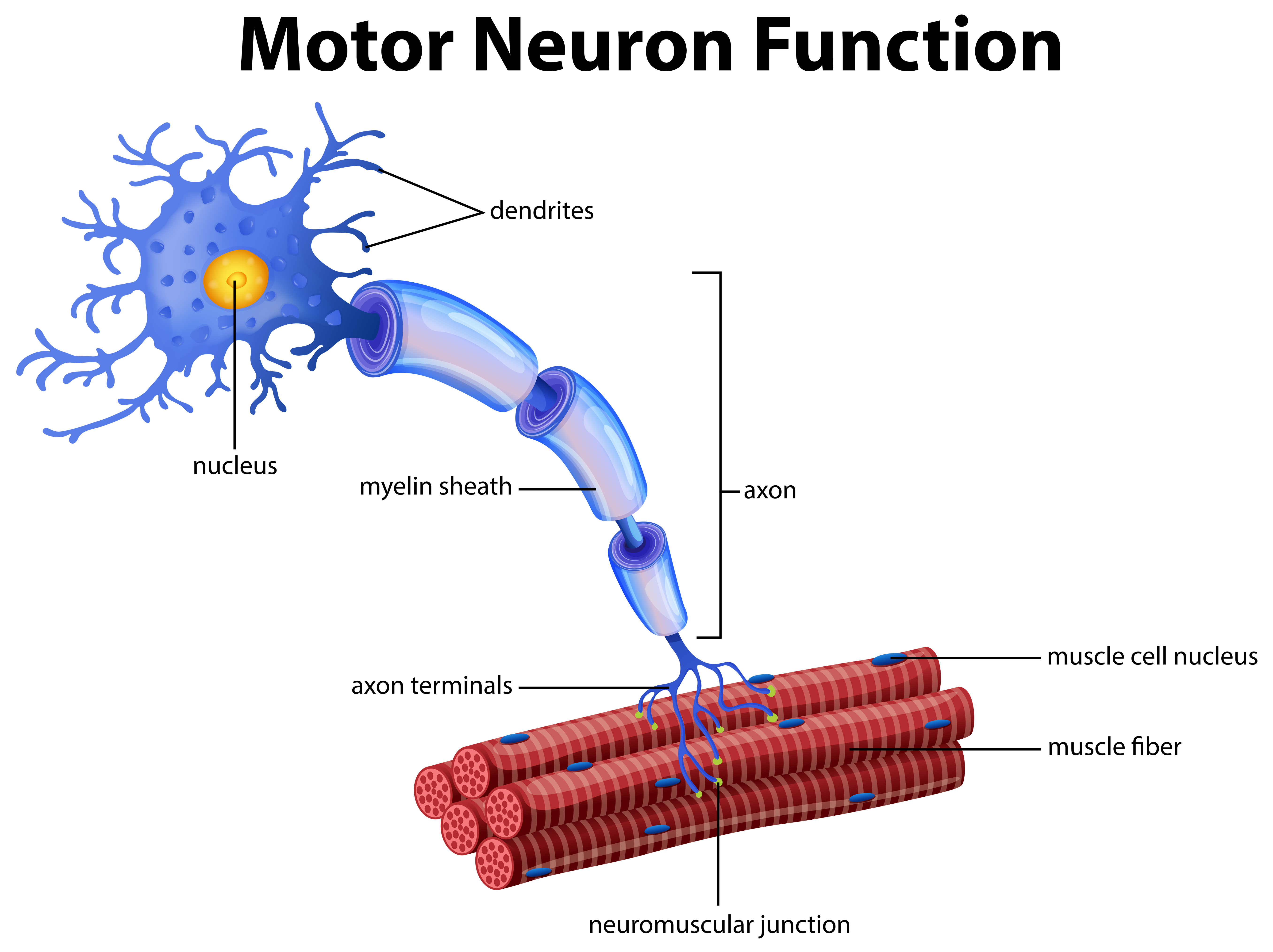
The target of the upper motor neuron is the dendrites of the lower motor neuron in the gray matter of the spinal cord. (8) The axon of the lower motor neuron emerges from the spinal cord in a nerve and connects to a muscle through a neuromuscular junction to cause contraction of the target muscle.

Structure of a motor neuron. Anatomy of a typical human neuron
Amyotrophic Lateral Sclerosis and Other Motor Neuron Diseases. 2023 Oct 1;29 (5):1538-1563. doi: 10.1212/CON.0000000000001345. This article reviews the clinical spectrum of amyotrophic lateral sclerosis (ALS), its variant presentations, and the approach to diagnosis and management. This review includes a detailed discussion of current and.
Motor Neuron The Definitive Guide Biology Dictionary
Essentially, motor neurons, also known as motoneurons, are made up of a variety of intricate, finely tuned circuits found throughout the body that innervate effector muscles and glands to enable both voluntary and involuntary motions. Two motor neurons come together to form a two-neuron circuit.

Neurons What are they and how do they work?
NIH HHS USA.gov While the term "motor neuron" evokes the idea that there is only one type of neuron that conducts movement, this is far from the truth.
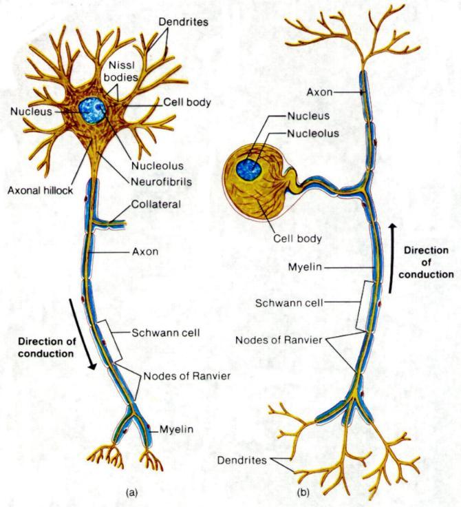
Histology of the Nervous System (The Neuron) Part 1
A motor neuron is a cell of the central nervous system. Motor neurons transmit signals to muscle cells or glands to control their functional output. When these cells are damaged in some way, motor neuron disease can arise. This is characterized by muscle wasting (atrophy) and loss of motor function. Motor Neuron Overview

Figure 7 4 Structure Of A Typical Motor Neuron Bangmuin Image Josh
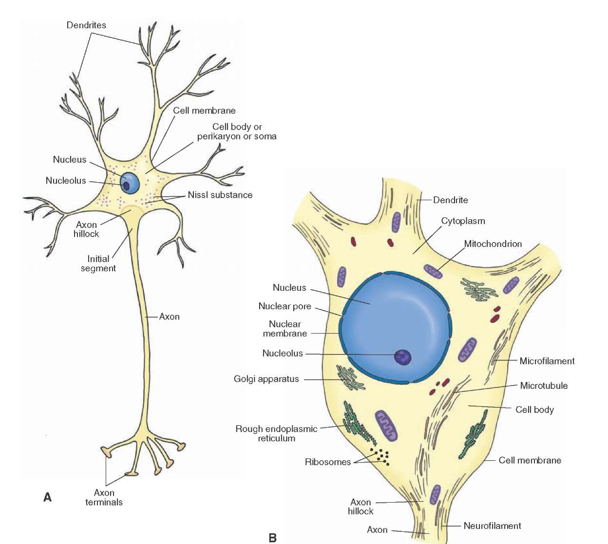
Neuron Structure. Figure \(\PageIndex{2}\) shows the structure of a typical neuron. The main parts of a neuron are labeled in the figure and described below. Figure \(\PageIndex{2}\): Somatic Motor Neuron with cell body, axon, axon, myelin sheath, nodes of Ranvier, axon terminal, dendrites, synaptic end of the bulbs, and other associated.

The Nervous System (Structure and Function) (Nursing) Part 1
A single oligodendrocyte can extend to up to 50 axons, wrapping around approximately 1 mm of each and forming the myelin sheath; Schwann cells, on the other hand, can only wrap around 1 axon. ( 27 votes) Upvote Flag
Motor neurons of the somatic nervous system pikolyourself
Well-Labelled Diagram of Motor Neuron A motor neuron is a nerve cell that functions to transmit signals from the central area of the nervous system to an effector site such as muscles or glands. A motor neuron can be broadly seen as consisting of three parts - cell body, axon and dendrites.
A Vector of Motor Neuron Function 296405 Vector Art at Vecteezy
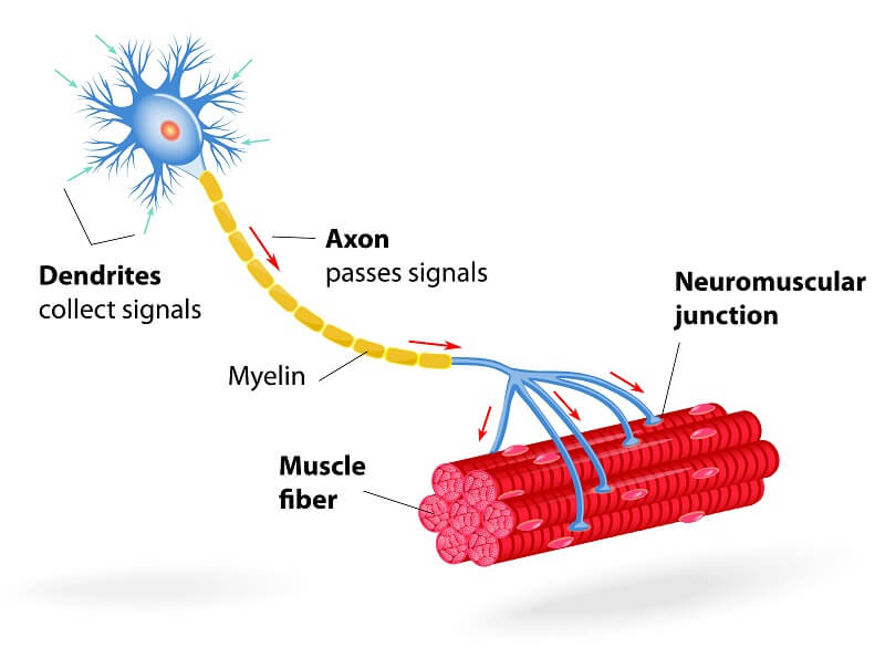
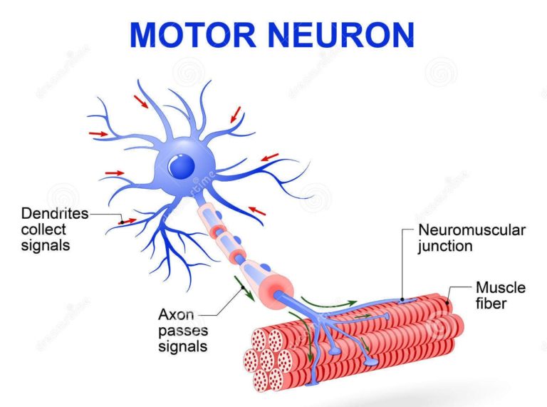
At its simplest, the neuromuscular junction is a type of synapse where neuronal signals from the brain or spinal cord interact with skeletal muscle fibers, causing them to contract. The activation of many muscle fibers together causes muscles to contract, which in turn can produce movement.

Neuroanatomy, Motor Neuron StatPearls NCBI Bookshelf
Sherrington was the first to recognize this fundamental relationship between an α motor neuron and the muscle fibers it innervates, for which he coined the term motor unit.Figure 16.4The motor unit. (A) Diagram showing a lower motor neuron in the spinal cord and the course of its axon to the muscle. (B) Each motor neuron synapses with multiple.